-1717753922-r.jpg)

Age-related macular degeneration (AMD) is a prevalent eye condition primarily affecting individuals over 50. It leads to a progressive deterioration of the retina's central portion, known as the macula. As the leading cause of vision loss among older adults, AMD presents a growing public health challenge, particularly in aging populations. Lets understand what makes this condition a priority challenge for the elderly and what can be done to manage it.
Defining Age-Related Macular Degeneration
Age-related macular Degeneration (AMD) is a prevalent eye condition primarily affecting older adults. It occurs when the macula, a small part of the retina responsible for clear central vision, deteriorates. This deterioration impacts daily activities such as reading, driving, and recognizing faces, leading to significant vision loss if not managed properly. AMD progresses differently among individuals, with some experiencing slow vision loss and others facing rapid and severe vision impairment.
Different Types Of Age-Related Macular Degeneration
Dry AMD
Dry AMD is the more common, accounting for about 80-90% of AMD cases. It results from gradually thinning the macula as part of the aging process. This type progresses slowly, causing mild to moderate vision loss. Drusen, yellow deposits of cellular debris beneath the retina are dry AMD. As these drusen grow more extensive and numerous, they can affect the retina's function, leading to vision problems.
Wet AMD
Wet AMD is less common but more severe. It occurs when abnormal blood vessels grow underneath the retina and leak blood and fluid. This leakage can cause rapid and significant damage to the macula, leading to sudden and severe vision loss. Wet AMD is responsible for most cases of advanced vision loss related to AMD. Prompt treatment is crucial to manage this type and preserve vision.
Recognizing Stages of Age-Related Macular Degeneration
Early AMD
In the early stage, individuals have medium-sized drusen but no noticeable vision loss. An eye exam can detect these changes even if the person is asymptomatic. Early detection is crucial for monitoring and managing the condition to prevent progression.
Intermediate AMD
Larger drusen and pigment changes in the retina mark intermediate AMD. Vision loss may start to occur, but it might not be significant enough to impact daily activities noticeably. Regular eye exams are essential at this stage to track progression and initiate preventive measures.
Late AMD
Late AMD involves noticeable vision loss and can be classified into geographic atrophy (dry AMD) and neovascular (wet AMD). Geographic atrophy refers to the thinning and losing the macula's tissues, while neovascular AMD involves abnormal blood vessel growth and leakage. Both forms significantly impact central vision, making early detection and treatment vital.
Major Symptoms of Age-Related Macular Degeneration

Blurred or Fuzzy Vision
Blurring or fuzzy vision is one of the earliest and most common symptoms of AMD. This symptom often affects the central field of vision, making it difficult to see fine details and perform tasks like reading or recognizing faces.
Difficulty Recognizing Faces
As AMD progresses, it can become increasingly challenging to recognize faces. This symptom is particularly distressing as it affects social interactions and the ability to identify loved ones.
Need for Brighter Light for Reading
Individuals with AMD often need brighter light for reading and other close-up tasks. This symptom results from the reduced ability of the macula to provide clear, detailed vision.
Difficulty Adapting to Low Light Levels
AMD can impair the eyes' ability to adjust to low light levels. This makes activities like driving at night or moving between differently lit environments challenging.
A Blurry or Blind Spot
A common sign of advanced AMD is the appearance of a blurry or blind spot in the center of the visual field. This spot can grow over time, significantly impairing central vision.
The Root Causes of Age-Related Macular Degeneration
Aging
The most significant risk factor for AMD is aging. As individuals age, the risk of developing AMD increases, particularly after the age of 50.
Oxidative Stress
Oxidative stress, caused by an imbalance between free radicals and antioxidants in the body, can contribute to AMD. This stress damages retinal cells, accelerating macular degeneration.
Inflammation
Chronic inflammation in the retina is believed to play a role in the development of AMD. Inflammatory processes can damage retinal tissues and contribute to disease progression.
Environmental Factors
Long-term exposure to harmful environmental factors, such as ultraviolet (UV) and blue light, can damage the retina and increase the risk of AMD. Protective eyewear and lifestyle adjustments can mitigate these risks.
Common Age-Related Macular Degeneration Risk Factors
Age
The risk of AMD increases significantly with age, particularly for individuals over 50. The likelihood of developing the condition continues to rise with advancing age.
Smoking
Smoking is a significant risk factor for AMD. Smokers are significantly more likely to develop the condition compared to non-smokers. Quitting smoking can reduce the risk and slow disease progression.
Check This Out: How Quitting Smoking Will Have Positive Effects
Race
Caucasians are at higher risk for AMD than other racial groups. Genetic and environmental factors may contribute to this increased susceptibility.
Obesity
A higher body mass index (BMI) increases the risk of AMD progression. Maintaining a healthy weight through diet and exercise can help reduce this risk.
Cardiovascular Disease
Conditions like hypertension and high cholesterol are linked to an increased risk of AMD. Managing cardiovascular health through medication and lifestyle changes can help mitigate this risk.
Effective Diagnosis of Age-Related Macular Degeneration

Visual Acuity Test
A visual acuity test measures the clarity of vision at various distances. It helps assess the impact of AMD on overall visual function.
Dilated Eye Exam
During a dilated eye exam, the ophthalmologist uses special drops to widen the pupil, allowing a thorough examination of the retina and macula. This exam can reveal drusen, pigment changes, and other signs of AMD.
Amsler Grid
The Amsler grid test is a simple tool for detecting central vision problems. Individuals with AMD may see distorted or missing lines on the grid, indicating macular damage.
Optical Coherence Tomography (OCT)
OCT is an imaging test that provides detailed cross-sectional images of the retina's layers. It helps detect and monitor changes in the macula, such as thinning or fluid accumulation.
Fluorescein Angiography
Fluorescein angiography involves injecting a fluorescent dye into the bloodstream and taking retina images. This test helps identify leaking blood vessels in cases of wet AMD.
Promising Treatment Options for Age-Related Macular Degeneration
Anti-VEGF Therapy
Anti-VEGF (vascular endothelial growth factor) therapy involves injecting medications into the eye to reduce the growth of abnormal blood vessels in wet AMD. These injections can help stabilize and improve vision.
Photodynamic Therapy
Photodynamic therapy uses a light-sensitive drug and a special laser to destroy abnormal blood vessels in the eye. This treatment is primarily used for wet AMD and can slow vision loss.
Laser Surgery
Though used less frequently, laser surgery can seal leaking blood vessels in wet AMD, helping prevent further damage to the macula.
Nutritional Supplements
The AREDS2 (Age-Related Eye Disease Study 2) formula, which includes vitamins C and E, zinc, copper, lutein, and zeaxanthin, has slowed the progression of intermediate and late AMD. These supplements support eye health and help delay vision loss.
Age-Related Macular Degeneration: Global Health Disparities
While AMD predominantly affects individuals of European descent, it's essential to recognize that this condition does not discriminate based on race or ethnicity. However, concerning trends have emerged regarding the diagnosis and treatment of AMD among different racial and ethnic groups. Recent studies have shed light on disparities in AMD care, revealing that Black patients are 23% less likely than their White counterparts to receive anti-VEGF injections, a primary treatment for wet AMD.
This treatment gap extends to Latino and Asian American individuals, who also face reduced access to essential therapies. Moreover, a significant discrepancy exists in the frequency of regular eye exams, a critical aspect of AMD management. Black individuals, in particular, are 18% less likely to undergo these exams compared to White individuals, leading to delayed detection and intervention.
Navigating the Challenges: Living with Age-Related Macular Degeneration
Management
Managing AMD involves regular monitoring and follow-up with an ophthalmologist. Early detection and timely intervention are critical to preserving vision. Vision aids, such as magnifiers and special glasses, can help individuals with AMD maintain their independence and quality of life.
Costs of Treatment
The costs of AMD treatment can vary widely, depending on the severity of the condition and the type of treatment required. Anti-VEGF injections, photodynamic therapy, and laser surgery can be expensive, but insurance coverage and financial assistance programs can help mitigate these costs. Additionally, nutritional supplements and vision aids may add to the overall expenses.
Lifestyle Changes
Adopting a healthy lifestyle can help manage AMD and reduce the risk of progression. A diet rich in leafy greens, fish, and nuts provides essential nutrients that support eye health. Regular exercise, maintaining a healthy weight, and avoiding smoking and excessive alcohol consumption can also benefit individuals with AMD.
Mental Preparedness
Coping with vision loss can be challenging and may require mental and emotional support. Joining support groups, seeking counseling, and engaging in activities that promote mental well-being can help individuals adjust to life with AMD. Staying informed about the condition and treatment options can empower individuals to manage their eye health actively.
Macular Degeneration Prognosis
The prognosis for AMD varies depending on the type and stage of the condition. While vision loss can be significant, many individuals retain some degree of vision, especially if diagnosed and treated early. Ongoing research and advancements in treatment continue to improve outcomes for those with AMD. Early detection, regular monitoring, and adherence to treatment plans are essential for preserving vision and maintaining quality of life.
Necessary Prevention of Age-Related Macular Degeneration
Quit Smoking
Quitting smoking is one of the most effective ways to reduce the risk of AMD. Smoking cessation can slow the progression of the disease and improve overall eye health.
Protect Eyes from UV Light
Wearing sunglasses that block UV rays can help protect the eyes from harmful sunlight. This preventive measure can reduce the risk of AMD and other eye conditions.
Maintain a Healthy Diet
A diet rich in antioxidants and omega-3 fatty acids supports eye health and can help prevent AMD. Foods such as leafy greens, fish, nuts, and fruits provide essential nutrients that protect the macula.
Check This Out: 20 Healthful Herbs You Must Add to Your Diet
Regular Exercise
Regular physical activity promotes overall health, including eye health. Exercise helps maintain a healthy weight and reduces the risk of AMD-related cardiovascular diseases.
Routine Eye Exams
Regular eye exams are crucial for early detection and management of AMD. Comprehensive eye exams can identify changes in the retina and macula before symptoms become apparent, allowing for timely intervention.
Conditions That May Mimic Age-Related Macular Degeneration
For esatblishing the correct diagnosis, it is crucial to investigate conditions that show similar signs and symptoms as that of AMD. Such as:
Diabetic Retinopathy
Diabetic retinopathy is a complication of diabetes that affects the blood vessels in the retina. It can cause vision loss like AMD, but the underlying cause is different.
Retinal Detachment
Retinal detachment occurs when the retina separates from the underlying tissue, leading to vision loss. Symptoms can include sudden flashes of light and floaters, which differ from AMD symptoms.
Central Serous Retinopathy
Central serous retinopathy involves fluid buildup under the retina, causing vision distortion. Unlike AMD, it primarily affects younger individuals and often resolves independently.
Retinal Vein Occlusion
Retinal vein occlusion occurs when a vein in the retina becomes blocked, leading to vision loss. This condition can mimic the symptoms of wet AMD but has different underlying causes.
Stargardt Disease
Stargardt disease is a genetic condition that causes progressive vision loss in younger individuals. It affects the macula similarly to AMD but occurs earlier in life.
Conclusion
Living with AMD requires proactive management and adaptation, but individuals can maintain a good quality of life with the right care and support. Regular eye exams, a healthy lifestyle, and staying informed about treatment options are crucial in managing this condition. By taking preventive measures and seeking timely treatment, individuals with AMD can preserve their vision and continue to lead fulfilling lives.
This content was created by AI
Recent Articles
What is Ehlers-Danlos Syndrome and How Can You Manage it?
Key TakeawaysEhlers-Danlos Syndrome is a group of inherited conditions that weaken the connective tissue holding your skin, joints, and organs together.There are 13 types, and the hypermobile and classical forms account for about 90% of diagnoses.Watch for very flexible joints, stretchy skin, easy bruising, frequent dislocations and joint pain that sticks around.There's no cure, but physical therapy, joint support and smart daily habits can ease symptoms and prevent complications.Sudden chest pain, fainting, vision changes or unexplained bleeding call for urgent medical care.Ever reached for a coffee cup and felt your shoulder slip out of place? For some people, that's just a normal Tuesday with Ehlers-Danlos Syndrome. The early signs are easy to wave off, which is part of the problem. Here's what the condition is, how it shows up, and when it's time to call a doctor.What is Ehlers-Danlos Syndrome?Ehlers-Danlos Syndrome, or EDS, is a group of inherited conditions that affect your connective tissues. Think of these tissues as the support system for your skin, joints, blood vessels, and many organs. Doctors recognize 13 types. The good news is that symptoms can usually be managed with the right plan.Ehlers-Danlos Syndrome Connective Tissue Disorder Explained SimplyConnective tissue works like the body's scaffolding, and it leans on two proteins, collagen and elastin. With Ehlers-Danlos Syndrome Connective Tissue Disorder, the collagen your body makes is weaker than it should be. Tissues end up loose or fragile. That's why symptoms can pop up in places that seem completely unrelated, from stretchy skin to loose joints.What are the Ehlers-Danlos Syndrome Types?Experts sort the condition into 13 types, and each one ties back to different genes. The Ehlers-Danlos Syndrome types vary in symptoms and risk, which is why an exact diagnosis matters so much. Five come up most often, so let's start there.Hypermobile EDSHypermobile EDS is the most common type. Your joints feel loose and unstable, and they may click, slip, or never quite seem to fit together. Doctors haven't tied it to one clear gene, so the diagnosis usually rests on your symptoms.Classical EDSClassical EDS looks a lot like the hypermobile type, but it hits the skin harder. Skin can feel soft and velvety, stretch easily and heal poorly, leaving thin, crinkly scars. Even stitches can tear out of fragile skin, so wound care deserves extra attention.Classical-Like EDSClassical-like EDS earns its name honestly. It closely resembles classical EDS, with only small differences in symptoms. That's why a provider has to review your history and examine your skin and joints before naming it. Getting the type right shapes how closely your team watches for related problems.Cardiac-Valvular EDSCardiac-valvular EDS affects the heart valves and cardiovascular system, along with the skin and joints. Valve problems can develop quietly, so regular heart checks matter for anyone with EDS. One of them is an echocardiogram, a noninvasive ultrasound that shows how your heart and valves look and work.Vascular EDSVascular EDS is very rare, but it's the most serious type because it involves the blood vessels. Vessel walls and organs like the intestines or uterus can tear without warning. If you're thinking about starting a family, experts recommend seeing a genetic counselor first, since pregnancy raises the risk of uterine rupture.What are the Ehlers-Danlos Syndrome Causes?The Ehlers-Danlos syndrome causes come down to genes. Changes in certain genes affect how your body builds and uses collagen, and researchers have found more than 20 of them. The specific change decides which type you get. Most types are inherited, so a parent can pass the change to a child. Sometimes it shows up for the first time with no family history at all. With hypermobile EDS, Mayo Clinic says each child has a 50% chance of inheriting it.What are the Common Ehlers-Danlos Syndrome Symptoms?Ehlers-Danlos syndrome symptoms look different from person to person. Still, loose joints, stretchy skin, and fragile skin show up in most types. Some people notice trouble as babies, while others don't see clear changes until much later. Let's look at adults and children separately.Ehlers-Danlos Syndrome Symptoms in AdultsEhlers-Danlos syndrome symptoms in adults often include joint pain, frequent dislocations, and a bone-deep tiredness that makes ordinary tasks harder. Easy bruising, hernias, irritable bowel syndrome, brain fog and trouble controlling urine or stool can join the list. Mayo Clinic also notes that very flexible joints can raise the risk of arthritis earlier in life.Ehlers-Danlos Syndrome Symptoms in ChildrenEhlers-Danlos syndrome symptoms in children usually begin in childhood, and some types appear as early as infancy. Parents might spot very bendy joints, soft stretchy skin, easy bruises, unusual scars, or joints that slip out of place. Because some types carry serious risks, an early evaluation gives your family time to build a safe plan.What are the Early Signs of Ehlers-Danlos Syndrome?Signs of Ehlers-Danlos Syndrome are easy to brush off, especially when they creep in slowly or look like plain clumsiness. Pay attention if several show up together, or if a relative already has a diagnosis.Joints that bend far past the normal range, click often, or slip out of place during simple movements.Soft, velvety skin that stretches and snaps back when you gently pinch itFragile skin that bruises easily, heals poorly and leaves thin, crinkly scarsOngoing joint or muscle pain, plus tiredness and brain fog with no clear causeHow Does Ehlers-Danlos Syndrome Diagnosis Work?Ehlers-Danlos syndrome diagnosis usually begins with a physical exam and a long conversation about your symptoms and medical history. Your provider will check your skin and joints, so tell them when things started and what makes them worse. Some types link to specific genes. Hypermobile EDS doesn't, so symptoms usually guide that diagnosis. Ask your provider to explain your exact type, because it shapes your care plan and screening schedule.What are the Ehlers-Danlos Syndrome Complications?Ehlers-Danlos syndrome complications depend on your type and symptoms, so risks vary a lot. Dislocations are the most common. Some problems are mild, while a few rare ones are serious and need quick action. The table below shows what doctors keep an eye on.ComplicationWhat It InvolvesJoint dislocationsLoose joints slip out of place and may need emergency care.Early arthritisVery flexible joints raise the risk of arthritis at a younger age.Heart valve problemsSome types affect the heart, so echocardiograms help catch changes early.Scoliosis and thin corneasSpine curves and eye changes can appear and need monitoring.Blood vessel ruptureRare vascular EDS can cause sudden, life-threatening internal bleeding.Must Read: Holistic Solutions for Amyloid Cardiomyopathy TreatmentWhat are the Ehlers-Danlos Syndrome Treatment Options?There's no cure, but Ehlers-Danlos syndrome treatment options aim to ease symptoms and prevent complications. Your plan will depend on your type, your symptoms, and your daily routine. And yes, most people can stay active once they learn which movements protect their joints and which ones cause strain.Physical Therapy and Joint BracesPhysical therapy builds the muscles around your joints, giving loose joints more support day to day. Braces can add stability where joints tend to slip. A good therapist will also teach you safe movement habits, so you get stronger without pushing joints past their comfortable range.Skin Protection and Gentle CareFragile skin needs gentle handling. Wear sunscreen and pick mild soaps. Ask your doctor how to care for wounds, since stitches in fragile skin can tear out and leave gaping scars. If you notice new bruising or bleeding that seems unusual, say so. It helps your provider adjust your plan.Smart Activity ChoicesYour provider may suggest skipping contact sports, high-impact exercise, and heavy lifting, since they raise the risk of dislocations. You can still move, though. You may just need to tweak things. A physical therapist can help you find low-impact activities that build muscle control without overloading your joints.Top Pick: When is the Best Time to Exercise for Peak Results?When Should You See a Doctor About Ehlers-Danlos Syndrome?Call your doctor if you notice loose joints with healing problems, dislocations, unexplained bruising, or a family history of EDS. Head to the emergency room right away if you or your child has any of these.Sudden pain or bleeding you can't explain, chest pain, or trouble breathingSudden dizziness or fainting when you stand upSudden vision changes, like silvery flashes or dark spots, which may signal a complicationConclusionEhlers-Danlos Syndrome has no cure, but early recognition and steady care can protect your joints, skin, and long-term health. Find out which type you have, stick with your care plan, and keep up with checkups so small problems get caught early. And if something feels off, trust your gut and talk to a provider who understands connective tissue conditions. This article offers general information only and isn't a substitute for medical advice.Frequently Asked QuestionsWhat is Ehlers-Danlos Syndrome in Simple Words?It's an inherited condition that weakens the connective tissue supporting your skin, joints, blood vessels and organs. Common signs include very loose joints, stretchy skin, fragile skin and a tendency to bruise easily.Can Ehlers-Danlos Syndrome Be Cured?No, there's no cure yet, but treatment can ease symptoms and lower the risk of complications. Physical therapy, joint braces, skin protection, and regular monitoring usually anchor a personal care plan for most people.Can Ehlers-Danlos Syndrome Be Inherited?Most types are inherited, so a parent carrying the gene change can pass it to a child. Sometimes the change appears for the first time, and genetic counseling can explain your family's chances.Are Ehlers-Danlos Syndrome Symptoms Different in Children?Symptoms often start in childhood, and some types appear in infancy while others show up much later. Tell your child's doctor early about loose joints, easy bruising, unusual scars or frequent joint dislocations.When Should I Go to the Emergency Room With Ehlers-Danlos Syndrome?Go for chest pain, trouble breathing, fainting, unexplained bleeding or sudden pain, and sudden vision changes. And never try to push a dislocated joint back into place yourself. Head to the emergency room.

What Causes Heart Palpitations and How are They Treated?
Quick TakeawayHeart palpitations create a sensation of fluttering, racing, or skipped beats in your chest or neck area.Common lifestyle triggers include high stress, excessive caffeine intake, dehydration, nicotine use, and intense physical exertion.Simple home techniques like deep breathing, vagal maneuvers, and drinking cold water can help slow down a racing pulse.Lying on your left side or eating heavy carbohydrate meals often increases awareness of your normal cardiac rhythm.Seek immediate medical evaluation if irregular heartbeat sensations occur alongside dizziness, fainting, or chest discomfort.Lying in bed at night and suddenly feeling your heart pound against your ribs can cause immediate worry. Experiencing heart palpitations makes you acutely aware of your heartbeat, creating uncomfortable sensations of fluttering, thumping, or skipping beats. According to clinical data published by the Cleveland Clinic, heart palpitations account for up to 16% of primary care medical visits and remain one of the most common reasons patients consult a cardiologist.While feeling an erratic rhythm can feel alarming, the vast majority of episodes are harmless responses to everyday stress, lifestyle factors, or hormonal shifts. Understanding what triggers these sensations empowers you to calm your nervous system and regain peace of mind. In this comprehensive medical guide, we will explore what causes fluttering feelings, examine common physical symptoms, discuss practical steps to stop episodes, and highlight red flag signs that indicate when to consult a healthcare provider.What is Heart Palpitation?Understanding what is heart palpitation starts with recognizing how your body senses cardiac activity. A palpitation is the conscious awareness of your own heartbeat. Under normal conditions, your heart pumps continuously without your conscious perception.During a palpitation episode, your heart may feel like it is beating too fast, pounding heavily, or skipping a beat entirely. These sensations can be felt in your chest, throat, or neck area. Although they feel unsettling, most sporadic episodes reflect benign extra heartbeats called premature atrial or ventricular contractions.What Do Heart Palpitations Feel Like, and What are Common Symptoms?Recognizing what do heart palpitations feel like helps individuals describe their exact physical symptoms to medical professionals accurately.Primary Heart Palpitations SymptomsCommon heart palpitations symptoms vary widely between individuals depending on the underlying trigger. Key sensations reported during an episode include:Fluttering: A gentle or rapid flapping sensation inside the chest cavity.Pounding: Heavy, forceful heartbeats that feel noticeable in the neck or ears.Skipped Beats: A brief pause in rhythm followed by a strong thumping heartbeat.Flip-Flopping: A sensation that the heart is physically turning over in the chest.Must Try: What is the Difference Between Heart Attack vs Heart FailureWhat are the Common Heart Palpitation Causes and Triggers?Identifying primary heart palpitations causes allows you to make simple daily adjustments that minimize future uncomfortable episodes.1. Emotional and Psychological StressWhen someone asks if anxiety can cause heart palpitations, the physiological answer is yes. Strong emotional stress, panic attacks, or fear trigger a surge of adrenaline in your bloodstream. This stress response elevates your heart rate and contracts blood vessels, directly causing fluttering feelings.2. Dietary and Lifestyle TriggersConsuming excessive stimulants like coffee, energy drinks, or nicotine alters electrical signaling in your cardiac tissue. Furthermore, experiencing heart palpitations after eating frequently occurs when digesting rich meals high in carbohydrates or sodium, which temporarily shifts blood flow toward the digestive tract.3. Nighttime and Positional ChangesIf you wonder why am I having heart palpitations at night, physical body position plays a major role. Experiencing heart palpitations when lying down on your left side compresses internal chest structures, making normal heartbeats resonate louder against your chest wall.How to Stop Heart Palpitations at Home?Learning how to stop heart palpitations quickly offers fast relief during sudden uncomfortable fluttering episodes.Management TechniqueHow It WorksBest Used ForDeep Belly BreathingActivates the parasympathetic nervous system to slow heart rateStress, anxiety, or panic-induced episodesVagal ManeuversStimulates the vagus nerve to regulate electrical cardiac signalsSudden rapid flutter sensationsHydration ResetRestores blood volume and balances vital electrolyte levelsDehydration or exertion triggersPracticing vagal maneuvers involves gently coughing, bearing down like a bowel movement, or splashing icy cold water onto your face. These physical actions stimulate your vagus nerve, which acts as a natural brake pedal for your cardiac electrical system.How Long Do Heart Palpitations Last, and Are They Dangerous?Understanding how long do heart palpitations last depends on the root trigger. Most harmless episodes caused by stress, caffeine, or sudden exertion resolve within a few seconds to a couple of minutes.When asking are heart palpitations dangerous, most isolated incidents in individuals without underlying cardiac disease are entirely benign. However, frequent episodes lasting several hours can indicate an underlying arrhythmia, thyroid disorder, or structural heart issue that requires professional medical assessment.When to See a Doctor for Heart Palpitations?Knowing when to see a doctor for heart palpitations and when to worry about heart palpitations ensures timely medical intervention when necessary.You should schedule a routine appointment with a physician if fluttering sensations occur frequently, worsen over time, or disrupt your daily activities. Conversely, seek immediate emergency medical evaluation if your palpitations are accompanied by:Chest Pain or Pressure: Uncomfortable tightness spreading to your arm, jaw, or back.Shortness of Breath: Difficulty catching your breath while resting.Dizziness or Fainting: Feeling lightheaded or losing consciousness suddenly.Pre-Existing Heart Conditions: A personal history of heart failure or valve disease.Try This: Types of Heart Attack: STEMI, NSTEMI, and Coronary SpasmWhat are the Medical Diagnosis and Heart Palpitations Treatment Options?Evaluating persistent cardiac symptoms involves comprehensive testing to determine proper heart palpitations treatment.Physicians typically conduct an electrocardiogram (ECG) to monitor real-time electrical activity. If episodes occur unpredictably at home, your doctor may recommend wearing a portable Holter monitor or patch for several days to capture elusive irregular rhythms. Treatment strategies focus on avoiding identified triggers, managing stress through mindfulness, adjusting medications, or utilizing prescribed beta-blockers to regulate electrical impulses when needed.ConclusionUnderstanding heart palpitations helps remove the anxiety and fear surrounding sudden chest fluttering or pounding sensations. Throughout this guide, we explored primary physical symptoms, identified common lifestyle triggers like stress and dietary habits, reviewed immediate home calming techniques, and outlined clear medical guidelines detailing when to seek professional care. While most palpitations represent harmless responses to daily stressors or body positioning, paying attention to your heart rhythm remains an essential component of personal wellness. Taking proactive steps to manage stress, stay hydrated, and maintain open communication with healthcare professionals keeps your heart beating smoothly for years to come.Are you ready to take control of your cardiovascular wellness and gain complete peace of mind? Connect with trusted medical insights and explore expert health resources today!FAQsWhat electrolyte deficiencies cause heart palpitations?Deficiencies in essential electrolytes like potassium, magnesium, calcium, and sodium frequently trigger heart palpitations. These minerals maintain proper electrical charges across cardiac muscle cells. Low levels disrupt normal signal transmission, causing premature heartbeats, muscle cramps, and erratic cardiac rhythms that require dietary adjustments or oral supplementation under medical supervision.Can hormonal changes during menopause trigger heart palpitations?Yes, hormonal fluctuations during perimenopause and menopause frequently cause heart palpitations. Decreasing estrogen levels affect the sympathetic nervous system and blood vessel elasticity, leading to sudden surges in heart rate. These episodes often accompany hot flashes or night sweats, resolving naturally as hormonal balance stabilizes over time.How does anemia contribute to heart palpitations?Anemia occurs when your body lacks sufficient healthy red blood cells to transport oxygen to vital tissues. To compensate for reduced oxygen levels, your heart must pump faster and harder to deliver necessary nutrients throughout the body, frequently resulting in noticeable pounding heartbeats, fatigue, and persistent shortness of breath.Does sleep apnea increase the risk of heart palpitations?Undiagnosed sleep apnea significantly increases the risk of heart palpitations and irregular cardiac rhythms. Repeated pauses in breathing during sleep cause sudden drops in blood oxygen levels, triggering adrenaline surges and high blood pressure. This nighttime strain irritates heart tissue, leading to frequent nocturnal cardiac palpitations and long-term cardiovascular stress.Can thyroid disorders cause sudden heart palpitations?An overactive thyroid gland, known as hyperthyroidism, produces excess thyroid hormone that accelerates your body's overall metabolism. This surge speeds up cardiac electrical signaling, causing persistent fast heartbeats, sudden chest fluttering, unintended weight loss, heat sensitivity, and elevated anxiety levels that require specialized medical management and targeted prescription treatment.

What is Fabry Disease, and How is it Treated for Recovery?
Quick TakeawayFabry disease is a rare inherited metabolic disorder caused by mutations in the GLA gene on the X chromosome.Deficient alpha-GAL enzyme activity leads to a toxic buildup of fatty substances called globotriaosylceramide in body tissues.Early symptoms often emerge in childhood as burning hand or foot pain, skin rashes, and reduced ability to sweat.Left untreated, the progressive accumulation of lipids causes severe progressive kidney damage, heart failure, and stroke risks.Modern treatment strategies combine targeted enzyme replacement therapy, chaperone drugs, and proactive organ-specific medical care.Living with unexplained burning pain in your hands or feet can feel overwhelming and frightening. Many individuals spend years searching for answers to mysterious symptoms that doctors struggle to identify. Fabry disease is a rare, inherited genetic condition that affects multiple organ systems throughout the body. According to clinical data from the National Institutes of Health, Fabry disease affects approximately 1 in 40,000 to 60,000 males worldwide, though milder late-onset forms occur even more frequently.Receiving a rare disease diagnosis often brings a mixture of relief and uncertainty about what the future holds. Understanding how this condition progresses helps individuals and families take proactive control of their health journey. In this medical guide, we will examine the biological causes, spot early warning signs in children and adults, discuss symptoms of Fabry disease, fabry disease causes, explain diagnostic testing, and discuss modern therapies designed to protect major organ function.What is Fabry Disease?Fabry disease is an inherited lysosomal storage disorder characterized by the abnormal accumulation of a specific type of fat called globotriaosylceramide (Gb3 or GL-3) in cells throughout the body. Because the body lacks sufficient active enzymes to break down these fatty substances, lipids build up continuously over time inside blood vessels, smooth muscle tissue, and major nerve networks.This gradual lipid buildup progressively impairs blood flow and compromises major organ systems. Left unmanaged, the condition damages blood vessels in the kidneys, heart, brain, and skin. While it is a lifelong progressive condition, early clinical identification allows medical specialists to introduce targeted therapies that slow disease progression and protect vital organ function.What Causes Fabry Disease?To understand how this progressive illness develops, it helps to examine the biological factors and genetic mechanisms involved. Knowing what causes Fabry disease clarifies why lipid accumulation impacts so many organ systems over time.1. GLA Gene Mutations and Alpha-GAL DeficiencyThe primary cause of Fabry disease is a mutation in the GLA gene on the X chromosome. The GLA gene provides the genetic instructions for producing the lysosomal enzyme alpha-galactosidase A (alpha-G). Genetic researchers have identified over 1,000 distinct mutations in the GLA gene.Severe Mutations: Nonsense or frameshift mutations completely halt alpha-GAL enzyme production, causing the classic early-onset form of the disease.Partial Functional Mutations: Missense mutations may allow the body to produce partially working enzymes, resulting in milder or late-onset variants that primarily affect the heart or kidneys later in life.Lysosomal Lipid Accumulation: Without adequate active alpha-GAL enzymes, cellular lysosomes cannot break down complex glycolipids like globotriaosylceramide, leading to toxic intracellular accumulation across blood vessel walls.2. X-Linked Genetic TransmissionThe underlying inheritance pattern of the disorder is directly linked to sex chromosomes:Transmission in Males: Males possess one X and one Y chromosome. Inheriting a mutated GLA gene on their single X chromosome severely reduces or eliminates alpha-GAL activity, triggering classic disease manifestations.Transmission in Females: Females possess two X chromosomes. A female with one mutated GLA gene is a carrier, but because of random X-chromosome inactivation (lyonization), her symptoms can range from completely asymptomatic to as severe as those in affected males.Must Try: Natural Remedies for Cardiac Amyloidosis: Home Care GuideWhat are the Common Symptoms of Fabry Disease?Recognizing early warning signs allows patients to seek medical intervention before major organs experience permanent structural damage. Fabry disease symptoms vary significantly based on biological sex, age, and individual genetic variation.Systemic Progression OverviewChildhood and Early Stages: Symptoms typically begin with episodes of burning limb pain (acroparesthesia) and reduced sweating (hypohidrosis).Adulthood Manifestations: As fat deposits grow in blood vessels, dark red skin spots (angiokeratomas) and persistent gastrointestinal distress become more pronounced.Late-Stage Complications: Chronic cellular accumulation eventually leads to progressive kidney impairment, cardiac enlargement, elevated risk of stroke, and potential organ failure.Fabry Disease in ChildrenInitial clinical signs usually emerge during childhood or adolescence between ages three and ten:Acroparesthesia: Episodes of severe burning or tingling pain in the hands and feet, often triggered by heat, exercise, stress, or fever.Hypohidrosis: A reduced ability to sweat, leading to severe heat intolerance, exercise avoidance, and sudden physical fatigue.Gastrointestinal Distress: Frequent stomach cramps, nausea, diarrhea, and abdominal pain shortly after eating meals.Fabry Disease in AdultsIf left unmanaged, fatty deposits continue accumulating across major vascular networks. Fabry disease in adults often presents with serious multi-organ complications:Organ SystemClinical ComplicationTypical Signs & ImpactsDermatologicalAngiokeratomasSmall, dark red or purple skin spots between the belly button and kneesOphthalmologicalCornea VerticillataWhorl-like clouding patterns on the cornea that do not impair visionRenalProgressive Kidney DamageProtein in the urine, reduced filtration, and eventual kidney failureCardiovascularHeart DiseaseEnlarged heart muscle, irregular heartbeats, and elevated stroke risksHow is Fabry Disease Diagnosed?Obtaining an accurate diagnosis requires specialized laboratory evaluations and genetic analysis. Doctors evaluate clinical history alongside targeted laboratory testing to confirm the diagnosis.Laboratory Blood and Genetic TestsUnderstanding how is Fabry disease diagnosed depends heavily on biological sex:Male Diagnostic Testing: An enzyme assay test measures alpha-GAL enzyme activity in white blood cells. Near-zero enzyme activity confirms a diagnosis.Female Diagnostic Testing: Females often show normal enzyme levels in blood samples. Molecular genetic testing must be performed to sequence the GLA gene and identify specific mutations.Prenatal and Family ScreeningOnce an individual receives a formal fabry disease diagnosis, physicians recommend family pedigree screening. Testing immediate family members helps identify asymptomatic relatives who carry the mutated gene early in life.What are the Modern Treatment Options for Fabry Disease?While there is currently no cure, advanced treatment options for Fabry disease focus on replacing missing enzymes, slowing disease progression, and managing specific organ symptoms.Targeted Medical TherapiesPrimary fabry disease treatment aims to clear cellular fat buildup and restore metabolic balance:Enzyme Replacement Therapy (ERT): Biweekly intravenous infusions deliver synthetic alpha-GAL enzymes directly into the bloodstream to reduce cellular lipid accumulation.Oral Chaperone Therapy: Oral medications assist specific unstable mutated enzymes to fold correctly, allowing them to function properly inside cells.Symptom and Organ SupportSecondary management relies on proactive organ care:Pain Management: Prescription medications help manage nerve-burning sensations and acroparesthesia spikes.Kidney and Heart Care: Blood pressure medications protect kidney filtration membranes and reduce strain on heart muscle walls.Try This: What are Infectious Diseases, and How are They Treated?ConclusionLiving with a rare genetic condition requires ongoing medical monitoring, personalized therapies, and strong educational resources. Throughout this guide, we explored how GLA gene mutations cause fatty accumulations, recognized key early warning signs in children and adults, examined genetic diagnostic methods, and detailed modern disease-modifying treatments. Early detection plays a vital role in preventing severe renal and cardiovascular complications. Working closely with geneticists, nephrologists, and cardiologists ensures you receive a comprehensive care plan tailored to your health goals.If you or a family member are navigating unexplained metabolic symptoms, take charge of your long-term health today. Speak with a specialized healthcare provider or genetic counselor to explore advanced diagnostic testing and modern treatment options!Frequently Asked QuestionsWhat life expectancy can someone with Fabry disease expect?Without treatment, Fabry disease can shorten life expectancy due to renal failure or heart complications. However, early diagnosis and modern disease-modifying therapies significantly improve life expectancy, allowing many treated individuals to live active, fulfilling lives into their sixties and beyond.How does Fabry disease affect the nervous system?Fabry disease damages small peripheral nerve fibers, causing burning pain in the hands and feet. Accumulating glycolipids in cerebral blood vessels also increases the risk of early-onset strokes, transient ischemic attacks, dizziness, and sudden hearing loss in affected adults.Can carrier females pass Fabry disease to their children?Yes, carrier females have a fifty percent chance of passing the mutated GLA gene to each child during pregnancy. Sons who inherit the mutation will develop the condition, while daughters who inherit the mutation will become carriers who may experience mild to severe symptoms.What lifestyle modifications help manage heat intolerance in Fabry disease?Patients managing heat intolerance should stay well hydrated, wear lightweight breathable clothing, and avoid strenuous exercise in hot weather. Using cooling vests, staying in air-conditioned environments, and taking cool baths help prevent painful acroparesthesia crises triggered by elevated body temperatures.Is gene therapy available as a treatment for Fabry disease?Gene therapy for Fabry disease is currently being evaluated in clinical research studies. Researchers are investigating viral vectors and gene editing techniques designed to instruct body tissues to produce functional alpha-GAL enzymes continuously, potentially eliminating the need for biweekly enzyme infusions in the future.
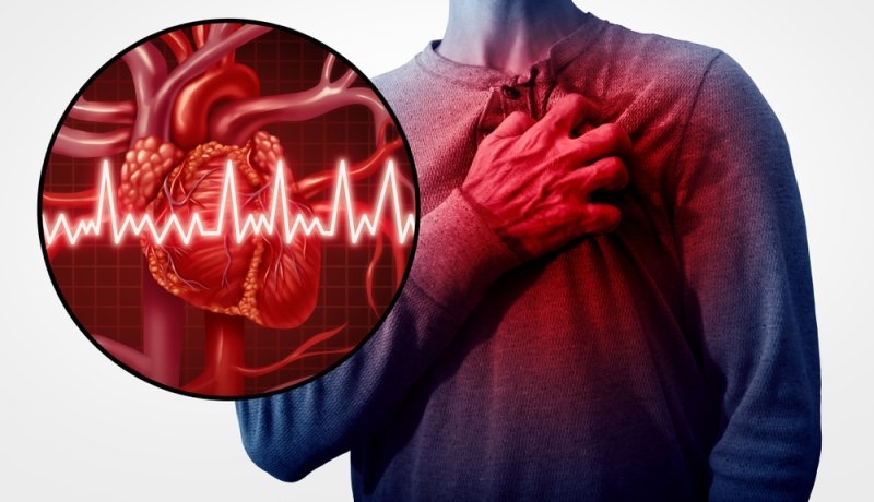
What is the Difference Between Heart Attack vs Heart Failure
Quick TakeawaysA heart attack is an acute circulation blockage, while heart failure is a chronic pumping weakness.Recognizing early warning signs of heart attack and heart failure can save lives during medical emergencies.Coronary artery disease is a leading cause for both conditions, making preventive care vital.Immediate emergency intervention restores blocked blood flow during a sudden cardiac event.Lifestyle modifications and medications help manage long-term cardiac function and overall health.Hearing a doctor mention cardiovascular disease can instantly cause panic and anxiety. Understanding heart attack vs. heart failure helps reduce fear by giving you clear, life-saving knowledge. You deserve accurate medical information to protect yourself and your loved ones during uncertain times.According to data from the Centers for Disease Control and Prevention, over 800,000 Americans experience a heart attack every year. Meanwhile, the American Heart Association reports that over 6 million adults in the United States currently live with heart failure. Knowing how these two cardiovascular events differ ensures you seek the right emergency care without dangerous delays.In this comprehensive guide, we will break down what is the difference between heart attack and heart failure, explore how is heart attack different from heart failure, examine heart attack vs. heart failure causes, and compare heart attack vs. heart failure treatment options. We will also answer critical questions like can a heart attack cause heart failure and evaluate heart attack or heart failure, which is more serious.What is a Heart Attack?A heart attack, medically known as a myocardial infarction, occurs when blood flow to a section of the heart muscle is suddenly blocked. This sudden blockage is usually caused by a buildup of plaque that ruptures and forms a blood clot in one of the coronary arteries. Without oxygen-rich blood, the affected heart muscle tissue begins to suffer damage and die rapidly unless emergency treatment quickly clears the blockage.What is Heart Failure?Heart failure is an ongoing condition in which the heart muscle becomes too weak or rigid to pump blood effectively throughout the body. Unlike a sudden heart attack, heart failure usually develops gradually over time due to chronic strain from conditions like high blood pressure or past cardiac damage. As pumping strength weakens, fluid can back up into the lungs, legs, and other tissues, leading to persistent fatigue and shortness of breath.How is Heart Attack Different From Heart Failure?Many people confuse these medical terms, but they represent entirely distinct cardiovascular problems.The core heart attack and heart failure difference lies in mechanism and speed. A heart attack is a sudden plumbing failure. A coronary artery gets blocked by plaque or a blood clot, starving the heart muscle of oxygen. Without prompt intervention, affected tissue dies quickly.In contrast, heart failure is a chronic pumping problem. The heart muscle becomes too weak or stiff to circulate enough blood to meet body needs. It develops slowly over months or years, causing fluid backup in the lungs and legs.What are the Warning Signs of Heart Attack and Heart Failure?Recognizing early symptoms ensures rapid medical evaluation and improves long-term recovery outcomes.Heart Attack SymptomsClassic heart attack symptoms appear suddenly and demand immediate emergency assistance.Crushing chest pain or pressure in the center of the chestPain radiating to the jaw, neck, back, or left armSudden shortness of breath accompanied by cold sweatsUnexplained dizziness, nausea, or extreme lightheadednessHeart Failure SymptomsProgressive heart failure symptoms build gradually over time as fluid accumulates in tissue.Chronic shortness of breath during routine physical activities or while lying flatPersistent swelling in the feet, ankles, legs, or abdomenConstant fatigue, weakness, and difficulty climbing stairsPersistent coughing or wheezing with white or pink blood-tinged phlegmHeart Attack vs Heart Failure Causes: What Triggers Each Condition?Understanding underlying risk factors helps patients take proactive steps toward prevention.Primary Heart Attack CausesMajor heart attack causes areCenter on plaque buildup inside arterial wallsHigh cholesterolSmokingHigh blood pressureDiabetes damages arterial linings over timeA ruptured plaque triggers rapid clot formation, completely blocking localized blood flowPrimary Heart Failure CausesCommon heart failure causes stem from chronic conditions that strain the cardiac muscle over time. Here are some other reasons:Long-standing hypertension forces the heart to pump against high pressureValvular heart diseaseCardiomyopathyThyroid disorders also weaken cardiac output gradually.Can a Heart Attack Cause Heart Failure?Yes, a severe cardiac event directly contributes to long-term pumping weakness. When a blocked artery starves heart tissue of oxygen during an acute event, those muscle cells die and form scar tissue. Scar tissue cannot contract or pump blood effectively. If a large portion of the heart muscle suffers damage, the remaining healthy tissue must work much harder. Over time, this extra burden leads directly to chronic heart failure.Must Try: What is Respiratory Syncytial Virus (RSV) and its Symptoms?Heart Attack vs Heart Failure Treatment: How are They Managed?Medical interventions focus on immediate stabilization for acute attacks and long-term management for chronic failure.Heart Attack Treatment ProtocolsEmergency heart attack treatment focuses on restoring blood flow immediately to minimize muscle death. Doctors use clot-dissolving medications or perform emergency angioplasty with stent placementCoronary artery bypass surgery may be performed in severe multi-vessel blockages.Heart Failure Treatment OptionsLong-term heart failure treatment aims to ease workload on the heart and control fluid buildup.Physicians prescribe ACE inhibitors, beta blockers, and diuretics to reduce blood pressure and swelling. Implantation of pacemakers or left ventricular assist devices helps stabilize advanced stages.Heart Attack or Heart Failure: Which is More Serious?Both conditions are severe, life-threatening medical issues that require professional cardiac care. A heart attack is an acute emergency where every minute determines muscle survival. Heart failure is a chronic condition that reduces life expectancy and daily quality of life. Neither condition should be ignored, as early intervention dramatically improves survival rates.ConclusionUnderstanding cardiovascular health allows you to make informed decisions for yourself and your family. In this guide, we explored key differences between acute arterial blockages and chronic pumping weakness. We examined distinct warning signs, underlying causes, and modern medical treatments available today. Recognizing early symptoms ensures you take action before permanent damage occurs. Consistent lifestyle choices like balanced nutrition, routine exercise, and regular blood pressure monitoring form your best defense. Taking control of your heart health today protects your vitality for decades to come. Are you ready to prioritize your cardiovascular wellness and gain complete peace of mind? Take the next step today by exploring our extensive medical library, connecting with qualified healthcare professionals, and discovering personalized wellness tools designed to support your lifelong vitality.Are you ready to take charge of your cardiovascular health and protect your future well-being? Connect with leading medical specialists today to access expert diagnostic guidance and personalized care plans tailored to your needs.FAQsWhat Medical Tests are Used to Diagnose Heart Attack vs. Heart Failure?Physicians use electrocardiograms and blood tests like troponin levels to diagnose an acute heart attack rapidly. To evaluate heart failure, doctors order echocardiograms to measure ejection fraction alongside BNP blood tests, which detect fluid overload and cardiac strain.How Does Ejection Fraction Measure Heart Function in Patients?Ejection fraction measures the percentage of blood the left ventricle pumps out with each contraction. A normal ejection fraction ranges between 50 and 70 percent. A lower reading indicates weakened pumping capacity characteristic of systolic heart failure.Can Lifestyle Modifications Reverse Early Stages of Heart Failure?While damaged heart tissue cannot fully regenerate, adoption of a low-sodium diet, regular exercise, and smoking cessation significantly improves heart function. These positive habits reduce cardiac strain and help manage symptoms effectively over time.What is Cardiac Rehabilitation and Who Needs it Most?Cardiac rehabilitation is a medically supervised program offering exercise training, emotional support, and lifestyle education. It benefits patients recovering from heart attacks, heart surgery, or heart failure by improving physical endurance and reducing future cardiovascular risks.How Do Diuretics Help Patients Manage Heart Failure Symptoms?Diuretics prompt the kidneys to remove excess water and sodium from the bloodstream through urination. Reducing fluid volume lowers blood pressure and relieves swelling in the legs, abdomen, and lungs, making breathing much easier for patients.

Types of Heart Attack: STEMI, NSTEMI, and Coronary Spasm
Key TakeawaysThe classification of different types of heart attacks depends on changes in blood flow and injuries sustained by the heart muscle, as well as ECG and blood test results.The fact is that STEMI and NSTEMI are myocardial infarction conditions, and coronary spasm is an acute condition as well if there is low blood flow.Similar symptoms can be observed when people experience different types of heart attacks, and such symptoms as chest pain, breathlessness, sweating, and nausea should be treated urgently.A heart attack, or, to put it in medical terms, myocardial infarction (MI), means a condition in which some part of the heart muscle lacks an oxygen-rich blood supply. The time for which the flow of blood has been reduced or blocked defines the chances of injury to the heart muscle. Since there can be different causes for heart attacks, the physician needs to evaluate the patient's symptoms, ECG, troponin tests, and imaging.Knowing the types of heart attacks can assist patients in having more information about their diagnosis. The basic types that are known to people are ST-segment elevation myocardial infarction (STEMI) and non-ST-segment elevation myocardial infarction (NSTEMI). Moreover, myocardial infarction or injury can also be caused by spasms of coronary arteries.What is a Heart Attack?A heart attack is when part of the heart muscle is cut off from blood. The most common cause is coronary artery disease, which is the buildup of plaque in arteries. The plaque can rupture, creating a clot that blocks the artery. Other less common reasons include constriction of the coronary artery or obstruction of blood flow.How are Heart Attacks Classified?The type of heart attack is determined by various factors other than the symptoms experienced. An electrocardiogram will indicate whether there is an ST-segment elevation or not. The presence of cardiac troponin, which is a protein found in heart cells, is measured using blood tests. Imaging will then be used to determine the affected artery and cause.STEMI and NSTEMI are two major categories of myocardial infarctions based on ECG findings. This does not imply that NSTEMI is a safe condition. Both need immediate medical attention. Moreover, the extent of damage to the heart muscle, the area where the block occurred, the presence of other health conditions, and the speed of treatment also have an effect on risk.Also check: Heart Attack Symptoms for Women: Common Signs and Risks STEMI: ST-Segment Elevation Myocardial InfarctionA STEMI heart attack is normally caused by the full blocking of a coronary artery, although there is no clear link between the two. Such disruption of the flow of blood could result in a significant injury to the heart muscles.It derives its name from the finding of an ST-segment elevation. Normally, the ECG is among the very first tests conducted when there is a suspected heart attack. In case of STEMI, the medical personnel usually work towards reopening the blocked coronary artery to restore the normal flow of blood.What is a STEMI Heart Attack?STEMI is a myocardial infarction that is characterized by ST-segment elevation in the ECG, and it is normally linked with an acute coronary artery blockage. Its symptoms include pain, squeezing or pressure in the chest, discomfort spreading to the arm, shoulder, back, neck, or jaw, shortness of breath, sweating, nausea, lightheadedness, or fatigue.Its treatment methods may include reopening the blocked artery via balloon angioplasty and stenting (PCI). Also, drugs that inhibit the formation of blood clots can be administered.NSTEMI: Non-ST-Segment elevation myocardial infarctionAnother type of myocardial infarction is non-ST-elevation myocardial infarction (NSTEMI). It does not cause ST-segment elevation that is characteristic of STEMI. But there is an elevation of cardiac troponins due to damage to myocardial tissue.What is an NSTEMI Heart Attack?A NSTEMI heart attack occurs when the blood flow to the coronary artery is severely restricted, most commonly because of a partial blockage of the vessel. Some patients experiencing NSTEMI can have a complete blockage of the artery, which is why just looking at the ECG cannot provide information about the whole picture.Symptoms of NSTEMI are similar to STEMI. Symptoms can include chest pain or discomfort, shortness of breath, nausea, sweating, dizziness, or pain or discomfort in the arms, back, neck, jaw, or upper stomach. The diagnosis is usually made based on symptoms and the patient's history, ECG results, and repeated measurement of cardiac troponin levels.Treatment depends on patient risk factors, extent of coronary disease, and other data. It includes anticoagulants/antiplatelet drugs, cholesterol-lowering therapy, and possibly coronary angiography followed by PCI.Coronary Artery SpasmThis is a process in which the artery suddenly tightens. This causes a temporary narrowing of the vessel, which causes a reduced flow of blood. It is also known as vasospastic or variant angina. Severe or persistent spasms may lead to myocardial infarction, even in the absence of fixed obstruction.What is a Coronary Spasm?Coronary spasm refers to a transient constriction of a coronary artery. The spasms occur during rest and are common during specific hours, especially overnight and in the morning hours. A key risk factor for spasms is smoking, whereas some individuals might have other triggers or accompanying conditions.Spasm does not imply the presence of a heart attack. If there is brief deprivation of blood flow that does not cause heart muscle death, the condition is categorized differently. A myocardial infarction develops if the lack of blood supply leads to myocardial damage.The symptoms of coronary spasm are similar to those of heart attacks. They include chest pain, profuse sweating, nausea, breathlessness, or faintness. Due to similarities, prompt evaluation is essential for proper diagnosis and treatment. Medications such as nitrates or calcium channel blockers may help reduce spasms.Symptoms of Heart Attacks Across Different TypesThe symptoms of different heart attacks may coincide; thus, you shouldn't try to determine the type of the disease using only its symptoms. Typical symptoms of a heart attack are the following:Pressure, tightness, squeezing, or pain in the chestPain extending to one's arms, shoulders, back, neck, or jawDifficulty breathingCold sweatsNauseaDizziness or faintingUnexpected fatigueSometimes there are very few symptoms, and one doesn't experience any chest pain. In case of a possible heart attack, you should call 911 (or a local emergency phone number) instead of trying to go to the hospital on your own.STEMI vs. NSTEMI vs. Coronary SpasmTypeCommon causeImportant diagnostic featurePrimary issueSTEMIUsually due to sudden blockage of coronary arteriesST segment elevation indicating injury to heart muscleVery quick heart muscle damageNSTEMIOften caused by severe restriction of coronary circulationTroponin increase but no diagnostic ST elevationInjury to heart muscleCoronary spasmSudden severe constriction of coronary arteriesDiagnosis through clinical evaluation, ECG changes, etc.Restriction of blood flow; sustained spasm leads to MI.This comparison explains why the terms are useful, but real cases can be more complicated. Doctors may use additional classifications based on the cause of the infarction, such as a supply-and-demand imbalance or another underlying condition.Which type of Heart Attack is more serious?This is impossible to know based on the information on the label. STEMI is usually viewed as the more acutely dangerous condition since there is usually a big obstruction present, and it can lead to a large amount of heart tissue damage if there is no prompt restoration of blood flow.NSTEMI is also dangerous and may indicate significant coronary heart disease. The outlook will depend on the extent of heart damage and where it occurs, what artery is involved, blood pressure and heart rate, kidney function, other medical problems, and the speed of treatment.The same goes for coronary artery spasm. An acute spasm may not do any lasting damage, while a more severe spasm may cause a myocardial infarction or serious rhythm disturbance.Must Read: Warning Signs of Blood Clotting Disorders: A GuideHow are Heart Attack types diagnosed and treated?The first tests that doctors usually run are the ECG and blood tests for cardiac troponin. Based on their results, they can employ echocardiography, coronary angiography, CT, or other imaging techniques. These procedures are used to assess whether a heart attack took place, whether there was any damage done, and whether it is necessary to restore an occluded artery.The choice of treatment methods depends on the type and cause of the heart attack. STEMI often requires immediate reperfusion, most frequently using PCI. NSTEMI may require drug therapy and coronary angiography, and coronary spasm may need relaxation medications.Despite the type, recovery often implies the administration of medications, participation in cardiac rehabilitation, follow-ups, and control of such factors as high blood pressure, high cholesterol, diabetes, smoking, diet, and exercise.ConclusionHeart attacks include types such as STEMI and NSTEMI; however, there is also coronary artery spasm, which can lead to myocardial infarction due to severe blood restriction. Even though the terms refer to different patterns and processes, all cases that could be heart attacks need urgent attention from medical professionals.Knowing the terminology will assist in understanding ECG findings, troponin tests, angiograms, and treatment plans; however, it must not stop you from calling for urgent medical assistance.Frequently Asked QuestionsCan there be a heart attack without having a blocked artery?Yes. A heart attack is possible even without complete blocking of an artery. A rupture of plaque, formation of a blood clot, severe constriction, and coronary artery spasms can result in insufficient blood flow to the heart muscle.When should one seek medical help in case of possible heart attack symptoms?Immediate medical assistance should be sought if the patient experiences symptoms like chest pressure, difficulty breathing, sweating, nausea, and others.Is it possible to have a heart attack with mild or unusual symptoms?Yes. There can be atypical signs of a heart attack, especially among older people and patients with particular diseases. It may be unusual tiredness, difficulty breathing, nausea, or pain in the neck, jaw, and arm.Will there be an increased risk for additional heart attacks after having a heart attack once?Previous history of a heart attack might increase the risks for subsequent heart problems. The use of ongoing treatments, therapies, and managing the risk factors could lower these risks.Can the damage done to the heart by a heart attack be undone?The damaged heart muscle tissue might be irreversible due to the death of cells from lack of blood flow. Nonetheless, fast action would ensure the restoration of blood flow and hence lessen the damage.
Popular Categories
Add Your Comment Here